Tag: Chronic lymphocytic leukaemia

  • Acute vs Chronic Leukaemia – A Brief Overview

    Acute vs Chronic Leukaemia – A Brief Overview

    Leukaemia is a group of malignancies affecting the bone marrow and blood, characterised by the uncontrolled proliferation of abnormal white blood cells. It is broadly classified into acute and chronic types, which differ significantly in presentation, pathophysiology, and management. This guide provides a structured overview to help junior doctors navigate the diagnosis and treatment of leukaemia.

    Classification of Leukaemia

    Leukaemias are divided based on cell lineage (myeloid vs lymphoid) and disease course (acute vs chronic):

    • Acute Myeloid Leukaemia (AML): Rapidly progressive cancer of myeloid progenitor cells.
    • Acute Lymphoblastic Leukaemia (ALL): Rapidly progressive cancer of lymphoid progenitor cells.
    • Chronic Myeloid Leukaemia (CML): Slow-growing cancer of myeloid cells.
    • Chronic Lymphocytic Leukaemia (CLL): Slow-growing cancer of lymphoid cells.

    Pathophysiology

    • Acute leukaemias arise from immature precursor cells (blasts), leading to uncontrolled proliferation and bone marrow failure.
    • Chronic leukaemias involve the accumulation of more mature but dysfunctional cells, leading to a slower disease progression.

    Clinical Presentation

    Acute Leukaemias (AML & ALL)

    • Rapid onset over weeks to months.
    • Symptoms of bone marrow failure, including anaemia (fatigue, pallor), neutropenia (infections), and thrombocytopenia (bleeding, bruising).
    • Systemic symptoms such as fever, weight loss, and night sweats.
    • ALL can involve the central nervous system (CNS), causing headaches, seizures, and neurological deficits.

    Chronic Leukaemias (CML & CLL)

    • Insidious onset over years, often detected incidentally on routine blood tests.
    • Fatigue, weight loss, and night sweats.
    • Lymphadenopathy and splenomegaly, especially in CLL.
    • CML may present with abdominal discomfort due to splenomegaly and a high white cell count.

    Investigations

    Acute Leukaemia

    • Full blood count (FBC): High or low white cell count with circulating blasts; anaemia and thrombocytopenia are common.
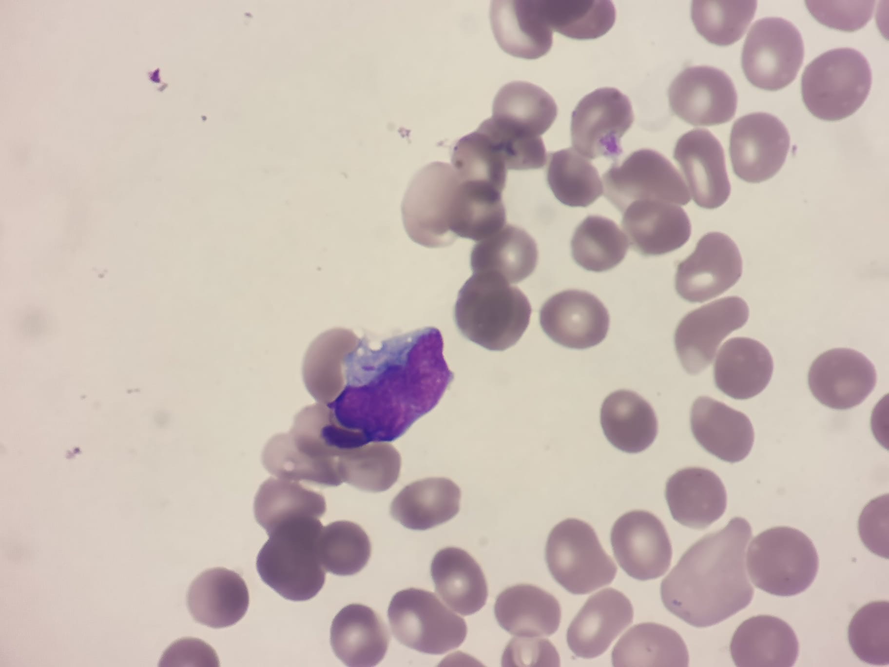
    • Blood film: Presence of blasts.
    • Bone marrow biopsy: Confirms diagnosis with >20% blasts.
    • Cytogenetics: Identifies translocations such as PML-RARA in acute promyelocytic leukaemia (APL).

    Chronic Leukaemia

    • FBC: Leukocytosis in CML; lymphocytosis in CLL.
    • Blood film: Smudge cells in CLL; left shift (immature myeloid cells) in CML.
    • Bone marrow biopsy: Hypercellular marrow with mature cells.
    • Cytogenetics: BCR-ABL fusion gene in CML.

    Management

    Acute Myeloid Leukaemia (AML)

    • Intensive chemotherapy with daunorubicin and cytarabine.
    • Bone marrow transplantation for high-risk or relapsed cases.

    Acute Lymphoblastic Leukaemia (ALL)

    • Multi-agent chemotherapy.
    • CNS prophylaxis with intrathecal methotrexate.
    • Bone marrow transplant for high-risk patients.

    Chronic Myeloid Leukaemia (CML)

    • First-line treatment with tyrosine kinase inhibitors (TKIs) such as imatinib or dasatinib.
    • Long-term suppression of disease with excellent survival rates.

    Chronic Lymphocytic Leukaemia (CLL)

    • Early-stage disease is often observed without treatment.
    • Symptomatic or high-risk disease is treated with targeted therapies like BTK inhibitors (ibrutinib) or BCL-2 inhibitors (venetoclax), often combined with monoclonal antibodies like rituximab or obinutuzumab.

    Prognosis

    • Acute leukaemias progress rapidly and require urgent treatment. Survival rates vary: AML has a 40-50% survival rate in younger patients but much lower in the elderly. ALL has a 70-90% survival rate in children but lower in adults.
    • Chronic leukaemias progress slowly and are often manageable for years. CML has an excellent prognosis with TKIs, with >90% five-year survival. CLL is highly variable, with some patients living decades without treatment.

    Key Takeaways for Junior Doctors

    • Acute leukaemias present with bone marrow failure and require urgent treatment.
    • Chronic leukaemias are often incidental findings and may not need immediate therapy.
    • Blasts in blood and bone marrow (>20%) suggest acute leukaemia.
    • CML is driven by BCR-ABL and treated with TKIs.
    • CLL is often indolent but requires treatment in high-risk cases.

    Leukaemia is a complex but fascinating field in haematology. Junior doctors play a vital role in recognising its presentation, initiating investigations, and understanding treatment pathways.

    Want to learn more? Join our in-depth haematology course at HaematologyForDoctors.com!

  • Chronic Lymphocytic Leukaemia (CLL) – A Junior Doctor’s Guide

    Chronic Lymphocytic Leukaemia (CLL) – A Junior Doctor’s Guide

    Chronic Lymphocytic Leukaemia (CLL) is the most common adult leukaemia in Western countries. It is a clonal disorder of mature B lymphocytes and has a highly variable clinical course, ranging from an indolent disease requiring no treatment to aggressive forms necessitating early intervention. This guide provides an overview of CLL for junior doctors rotating through haematology.

    Understanding CLL

    CLL is characterised by the accumulation of small, mature-looking but dysfunctional B lymphocytes in the blood, bone marrow, and lymphoid tissues. It is closely related to small lymphocytic lymphoma (SLL), with the distinction being that CLL primarily affects peripheral blood, whereas SLL involves lymphoid organs.

    Clinical Presentation

    CLL can be asymptomatic and detected incidentally on a routine full blood count. Symptomatic patients may present with:

    • Lymphadenopathy – Most common initial presentation.
    • Hepatosplenomegaly – Occurs in advanced disease.
    • Recurrent infections – Due to immune dysfunction and hypogammaglobulinaemia.
    • Fatigue & Weight Loss – B symptoms in more aggressive disease.
    • Autoimmune Complications – Autoimmune haemolytic anaemia (AIHA) and immune thrombocytopenia (ITP).

    Investigations

    • Full Blood Count (FBC) & Blood Film – Lymphocytosis (>5 × 10⁹/L) with small mature lymphocytes and smear (smudge) cells.
    • Flow Cytometry – Confirms clonal B-cell population with CD5, CD19, CD20 (dim), and CD23 positivity.
    • Bone Marrow Aspirate & Biopsy – Not always required but shows increased lymphocytes.
    • Cytogenetics & Molecular Testing – Prognostic markers:
      • FISH for del(13q), del(11q), del(17p) (p53 mutation – poor prognosis).
      • IGHV mutation status (mutated = better prognosis, unmutated = worse prognosis).
    • Immunoglobulin Levels – Assesses hypogammaglobulinaemia.

    Staging

    Two staging systems guide prognosis and treatment decisions:

    • Rai Staging (USA):
      • 0: Lymphocytosis only (low risk).
      • I-II: Lymphadenopathy and/or organomegaly (intermediate risk).
      • III-IV: Anaemia and/or thrombocytopenia (high risk).
    • Binet Staging (Europe):
      • A: <3 lymphoid areas involved.
      • B: ≥3 lymphoid areas involved.
      • C: Anaemia and/or thrombocytopenia.

    Management

    Management depends on disease stage, symptoms, and genetic markers. Many patients do not require immediate treatment and are monitored under a watch and wait approach.

    Supportive Care

    • Infection Prevention – Vaccination (e.g., pneumococcal, influenza), IV immunoglobulin for recurrent infections.
    • Autoimmune Cytopenias – Managed with steroids, rituximab, or immunosuppressants.
    • Blood Transfusions – Symptomatic anaemia or thrombocytopenia.

    Definitive Treatment

    1. Indications for Treatment
      • Symptomatic disease (B symptoms, bulky lymphadenopathy, organomegaly).
      • Progressive marrow failure (anaemia or thrombocytopenia).
      • Rapid lymphocyte doubling time (<6 months).
      • Severe autoimmune complications unresponsive to steroids.
    2. First-Line Therapy
      • Targeted Therapy (preferred):
        • Bruton’s tyrosine kinase (BTK) inhibitors: Ibrutinib, Acalabrutinib.
        • BCL-2 inhibitors: Venetoclax (often combined with anti-CD20 monoclonal antibodies like rituximab or obinutuzumab).
      • Chemoimmunotherapy (for selected fit patients with IGHV-mutated disease and no TP53 mutation):
        • FCR regimen (Fludarabine, Cyclophosphamide, Rituximab) – Younger patients.
        • Bendamustine + Rituximab – Older or frail patients.
        • Note this is (extremely) rarely used in the modern era
    3. Relapsed/Refractory CLL
      • BTK inhibitors (Ibrutinib, Acalabrutinib) or BCL-2 inhibitors (Venetoclax) if not used first-line.
      • PI3K inhibitors (e.g., Idelalisib) in selected cases.
      • Allogeneic stem cell transplantation for high-risk, refractory disease.

    Palliative Care

    • For elderly or frail patients with slow disease progression, best supportive care is often appropriate.
    • Symptom management with transfusions, steroids for autoimmune cytopenias, and infection prophylaxis.
    • Clear goals-of-care discussions, particularly in advanced disease.

    Complications to Watch For

    • Infections – Bacterial, viral (zoster), and fungal due to immunosuppression.
    • Tumour Lysis Syndrome (TLS) – Need to assess TLS risk and institute management as required when starting venetoclax.
    • Richter’s Transformation – Transformation to an aggressive lymphoma (diffuse large B-cell lymphoma); suspect in rapidly enlarging lymph nodes or new B symptoms.
    • Autoimmune Cytopenias – Haemolytic anaemia or thrombocytopenia.
    • Secondary Malignancies – Increased risk due to immune dysfunction and prior treatments.

    Prognosis and Follow-Up

    • Indolent cases may never require treatment and have a normal life expectancy.
    • Patients with TP53 mutations, del(17p), or unmutated IGHV have a poorer prognosis and require novel targeted therapies.
    • Regular follow-up includes monitoring blood counts, lymphadenopathy, and signs of transformation or progression.

    Key Takeaways for Junior Doctors

    1. CLL is often asymptomatic and detected incidentally.
    2. Flow cytometry confirms the diagnosis by demonstrating a clonal B-cell population.
    3. Many patients follow a watch and wait approach, with treatment initiated only when clinically necessary.
    4. Targeted therapies (BTK and BCL-2 inhibitors) have replaced chemotherapy for most patients.
    5. Recognising Richter’s transformation and autoimmune complications is crucial for prompt intervention.
    6. Palliative care should be considered for frail patients with advanced disease.

    CLL is a fascinating and heterogenous disease with a rapidly evolving treatment landscape. Junior doctors play an essential role in recognising its presentation, monitoring progression, and initiating appropriate management.


    Want to expand your haematology knowledge? Our course at HaematologyForDoctors.com covers real-world case discussions, treatment updates, and practical management strategies. Sign up today!