Tag: CML

  • Acute vs Chronic Leukaemia – A Brief Overview

    Acute vs Chronic Leukaemia – A Brief Overview

    Leukaemia is a group of malignancies affecting the bone marrow and blood, characterised by the uncontrolled proliferation of abnormal white blood cells. It is broadly classified into acute and chronic types, which differ significantly in presentation, pathophysiology, and management. This guide provides a structured overview to help junior doctors navigate the diagnosis and treatment of leukaemia.

    Classification of Leukaemia

    Leukaemias are divided based on cell lineage (myeloid vs lymphoid) and disease course (acute vs chronic):

    • Acute Myeloid Leukaemia (AML): Rapidly progressive cancer of myeloid progenitor cells.
    • Acute Lymphoblastic Leukaemia (ALL): Rapidly progressive cancer of lymphoid progenitor cells.
    • Chronic Myeloid Leukaemia (CML): Slow-growing cancer of myeloid cells.
    • Chronic Lymphocytic Leukaemia (CLL): Slow-growing cancer of lymphoid cells.

    Pathophysiology

    • Acute leukaemias arise from immature precursor cells (blasts), leading to uncontrolled proliferation and bone marrow failure.
    • Chronic leukaemias involve the accumulation of more mature but dysfunctional cells, leading to a slower disease progression.

    Clinical Presentation

    Acute Leukaemias (AML & ALL)

    • Rapid onset over weeks to months.
    • Symptoms of bone marrow failure, including anaemia (fatigue, pallor), neutropenia (infections), and thrombocytopenia (bleeding, bruising).
    • Systemic symptoms such as fever, weight loss, and night sweats.
    • ALL can involve the central nervous system (CNS), causing headaches, seizures, and neurological deficits.

    Chronic Leukaemias (CML & CLL)

    • Insidious onset over years, often detected incidentally on routine blood tests.
    • Fatigue, weight loss, and night sweats.
    • Lymphadenopathy and splenomegaly, especially in CLL.
    • CML may present with abdominal discomfort due to splenomegaly and a high white cell count.

    Investigations

    Acute Leukaemia

    • Full blood count (FBC): High or low white cell count with circulating blasts; anaemia and thrombocytopenia are common.
    • Blood film: Presence of blasts.
    • Bone marrow biopsy: Confirms diagnosis with >20% blasts.
    • Cytogenetics: Identifies translocations such as PML-RARA in acute promyelocytic leukaemia (APL).

    Chronic Leukaemia

    • FBC: Leukocytosis in CML; lymphocytosis in CLL.
    • Blood film: Smudge cells in CLL; left shift (immature myeloid cells) in CML.
    • Bone marrow biopsy: Hypercellular marrow with mature cells.
    • Cytogenetics: BCR-ABL fusion gene in CML.

    Management

    Acute Myeloid Leukaemia (AML)

    • Intensive chemotherapy with daunorubicin and cytarabine.
    • Bone marrow transplantation for high-risk or relapsed cases.

    Acute Lymphoblastic Leukaemia (ALL)

    • Multi-agent chemotherapy.
    • CNS prophylaxis with intrathecal methotrexate.
    • Bone marrow transplant for high-risk patients.

    Chronic Myeloid Leukaemia (CML)

    • First-line treatment with tyrosine kinase inhibitors (TKIs) such as imatinib or dasatinib.
    • Long-term suppression of disease with excellent survival rates.

    Chronic Lymphocytic Leukaemia (CLL)

    • Early-stage disease is often observed without treatment.
    • Symptomatic or high-risk disease is treated with targeted therapies like BTK inhibitors (ibrutinib) or BCL-2 inhibitors (venetoclax), often combined with monoclonal antibodies like rituximab or obinutuzumab.

    Prognosis

    • Acute leukaemias progress rapidly and require urgent treatment. Survival rates vary: AML has a 40-50% survival rate in younger patients but much lower in the elderly. ALL has a 70-90% survival rate in children but lower in adults.
    • Chronic leukaemias progress slowly and are often manageable for years. CML has an excellent prognosis with TKIs, with >90% five-year survival. CLL is highly variable, with some patients living decades without treatment.

    Key Takeaways for Junior Doctors

    • Acute leukaemias present with bone marrow failure and require urgent treatment.
    • Chronic leukaemias are often incidental findings and may not need immediate therapy.
    • Blasts in blood and bone marrow (>20%) suggest acute leukaemia.
    • CML is driven by BCR-ABL and treated with TKIs.
    • CLL is often indolent but requires treatment in high-risk cases.

    Leukaemia is a complex but fascinating field in haematology. Junior doctors play a vital role in recognising its presentation, initiating investigations, and understanding treatment pathways.

    Want to learn more? Join our in-depth haematology course at HaematologyForDoctors.com!
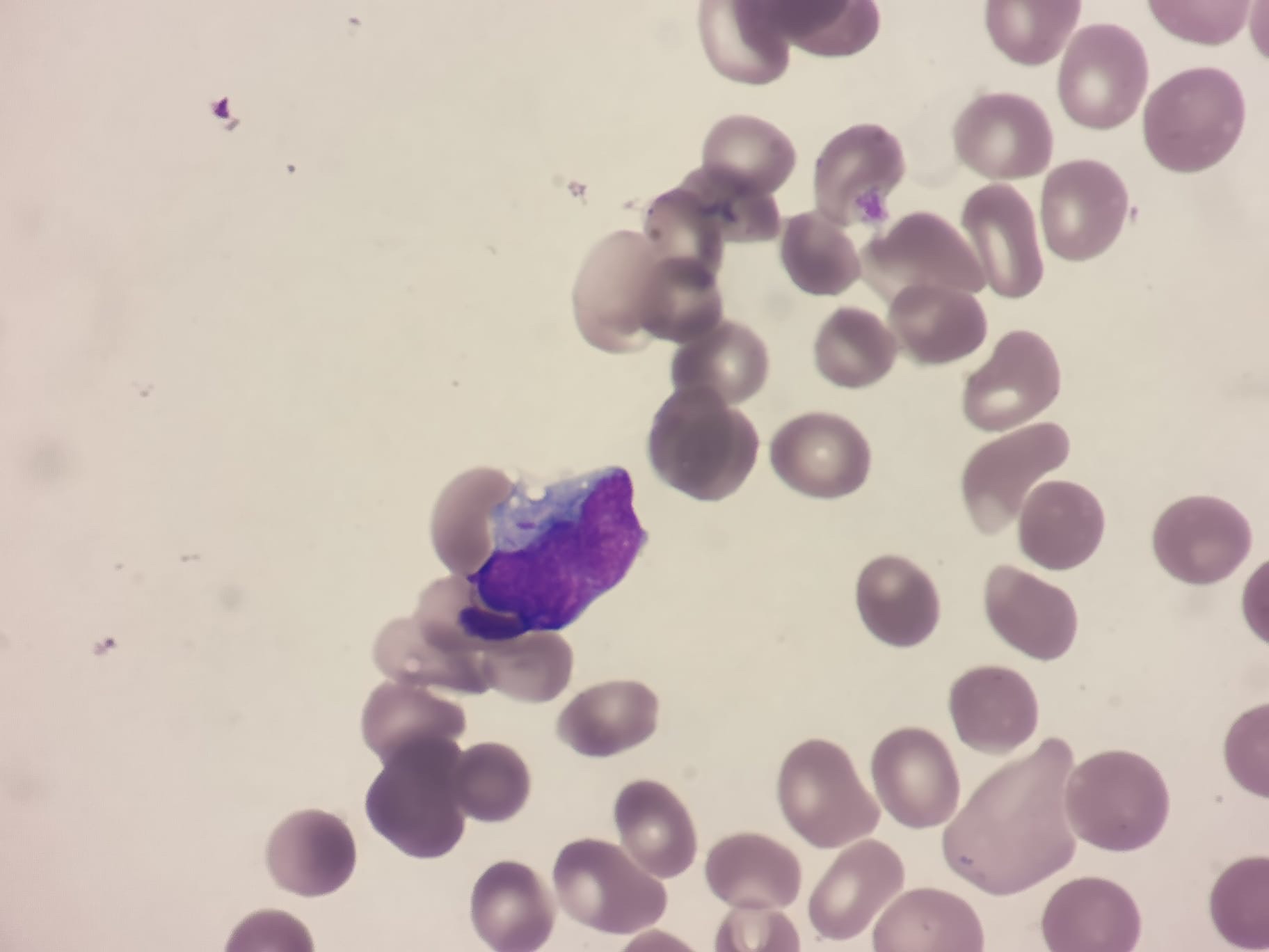
  • Chronic Myeloid Leukaemia (CML) – A Junior Doctor’s Guide

    Chronic Myeloid Leukaemia (CML) is a myeloproliferative disorder characterised by the uncontrolled proliferation of myeloid cells in the bone marrow. It is unique among leukaemias due to its strong association with a specific genetic abnormality, the Philadelphia chromosome (t(9;22)), which leads to the formation of the BCR-ABL fusion gene. This guide provides an overview of CML for junior doctors rotating through haematology.

    Understanding CML

    CML is a triphasic disease with distinct clinical phases:

    • Chronic Phase – Most patients are diagnosed in this phase, which is typically indolent and well-managed with targeted therapy.
    • Accelerated Phase – Increased disease activity with worsening cytopenias, rising white cell counts, and worsening symptoms.
    • Blast Crisis – Resembles acute leukaemia, with a poor prognosis unless treated aggressively.

    Clinical Presentation

    CML is often detected incidentally on routine blood tests. However, symptomatic patients may present with:

    • Fatigue and weight loss – Due to increased metabolic activity.
    • Splenomegaly – Leading to early satiety, left upper quadrant pain, or abdominal discomfort.
    • Night sweats and fever – Indicative of disease progression.
    • Bone pain – Due to increased myeloid proliferation.
    • Bleeding/bruising – If thrombocytopenia develops in later stages.

    Investigations

    A structured diagnostic approach is key when CML is suspected:

    • Full Blood Count (FBC) & Blood Film – Shows leucocytosis with a left shift (neutrophils, myelocytes, basophilia, and eosinophilia).
    • Bone Marrow Aspirate & Biopsy – Hypercellular marrow with increased granulopoiesis.
    • Cytogenetics & Molecular Testing –
      • Philadelphia chromosome detection via karyotyping.
      • BCR-ABL1 fusion gene confirmed via PCR or fluorescence in situ hybridisation (FISH).
    • Lactate Dehydrogenase (LDH) & Uric Acid – Markers of high cell turnover.

    Management

    Treatment of CML has been revolutionised by the introduction of tyrosine kinase inhibitors (TKIs), which specifically target the BCR-ABL fusion protein.

    Supportive Care

    • Hydroxyurea – Used for cytoreduction in patients with very high white cell counts at diagnosis.
    • Allopurinol & Hydration – To prevent tumour lysis syndrome (TLS).

    Definitive Treatment

    1. Tyrosine Kinase Inhibitors (TKIs) – The cornerstone of CML treatment, including:
      • First-line: Imatinib (first-generation), Dasatinib, Nilotinib (second-generation).
      • For resistant disease: Bosutinib, Ponatinib (third-generation).
      • TKI response monitoring: Measured via BCR-ABL1 transcript levels using quantitative PCR.
    2. Stem Cell Transplantation – Considered for TKI-resistant disease or blast crisis.
    3. Chemotherapy – Used in blast crisis to induce remission before transplant.

    Palliative Care

    • For patients who fail multiple lines of therapy or are unfit for aggressive treatment, supportive care focuses on symptom management and maintaining quality of life.
    • Palliative options include hydroxyurea for cytoreduction and symptom control.
    • End-of-life discussions and advanced care planning should be initiated when disease progresses despite therapy.

    Complications to Watch For

    • Disease Progression – Transition from chronic to accelerated phase or blast crisis.
    • TKI Resistance or Intolerance – Requires switching therapy or alternative treatment strategies.
    • Cardiotoxicity & Vascular Events – Seen with second- and third-generation TKIs.
    • Myelosuppression – TKI-induced cytopenias may require dose modifications.
    • Tumour Lysis Syndrome (TLS) – Can occur at treatment initiation, requiring prophylactic measures.

    Prognosis and Follow-Up

    • With TKIs, CML has transformed into a manageable chronic condition with near-normal life expectancy if well-controlled.
    • Regular monitoring with BCR-ABL1 PCR ensures response to therapy and detects early resistance.
    • Some patients in deep remission may attempt treatment-free remission (TFR) under strict monitoring.

    Key Takeaways for Junior Doctors

    1. CML is strongly linked to the Philadelphia chromosome (t(9;22)) and BCR-ABL fusion gene.
    2. Tyrosine kinase inhibitors (TKIs) have revolutionised treatment and significantly improved survival.
    3. Monitoring BCR-ABL1 transcript levels via PCR is crucial for assessing treatment response.
    4. Recognising disease progression from chronic phase to blast crisis is critical.
    5. Palliative care is an important option for patients who are refractory to treatment or unfit for aggressive therapy.

    CML is a fascinating disease that exemplifies the power of targeted therapy in modern haematology. Understanding its phases, management, and complications will help junior doctors provide optimal patient care.


    Want to learn more about haematological malignancies? Our haematology course provides in-depth case discussions, management strategies, and exam-focused content. Sign up now at HaematologyForDoctors.com!