Tag: junior doctor

  • The RATIFY Trial: Midostaurin in FLT3-Mutated AML

    The RATIFY Trial: Midostaurin in FLT3-Mutated AML

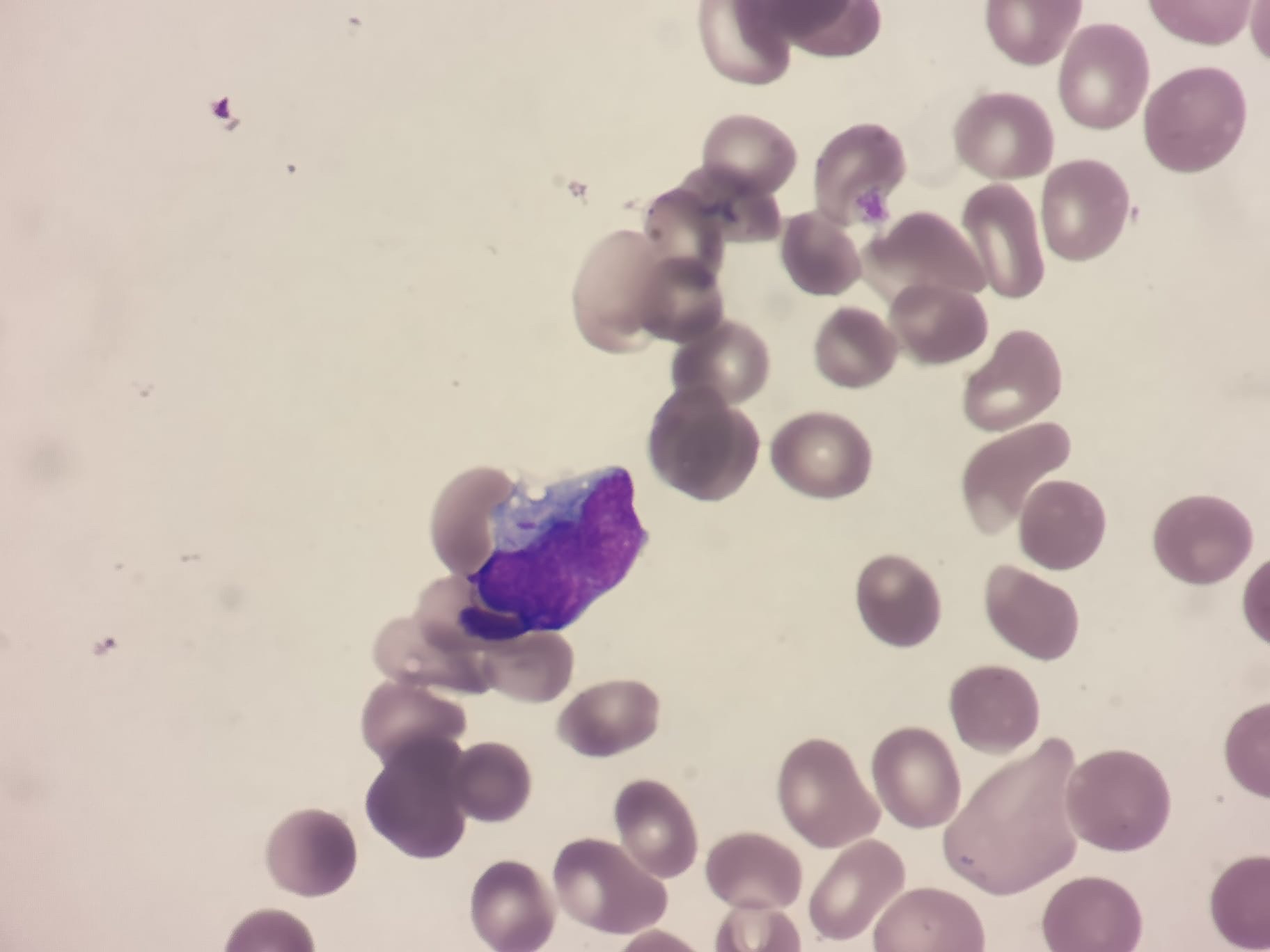
    Have you ever wondered why we give patients with FLT3 mutated AML midostaurin during their induction and consolidation chemotherapy? Today we will be talking about the RATIFY trial to find out.

    The RATIFY trial (CALGB 10603) was a pivotal phase III, randomized, double-blind, placebo-controlled study that evaluated the efficacy of adding midostaurin, a multikinase FLT3 inhibitor, to standard chemotherapy in adults aged 18–59 with newly diagnosed FLT3-mutated acute myeloid leukemia (AML). The trial enrolled 717 patients across 225 sites in 17 countries.

    Study Design

    • Population: Patients aged 18–59 with newly diagnosed AML harboring FLT3 mutations (including both internal tandem duplication [ITD] and tyrosine kinase domain [TKD] mutations).
    • Intervention: All patients received standard induction chemotherapy (daunorubicin and cytarabine). Those achieving remission proceeded to consolidation therapy. Patients were randomized to receive either midostaurin (50 mg twice daily) or placebo, administered during induction, consolidation, and as maintenance therapy for up to 12 months.

    Key Outcomes

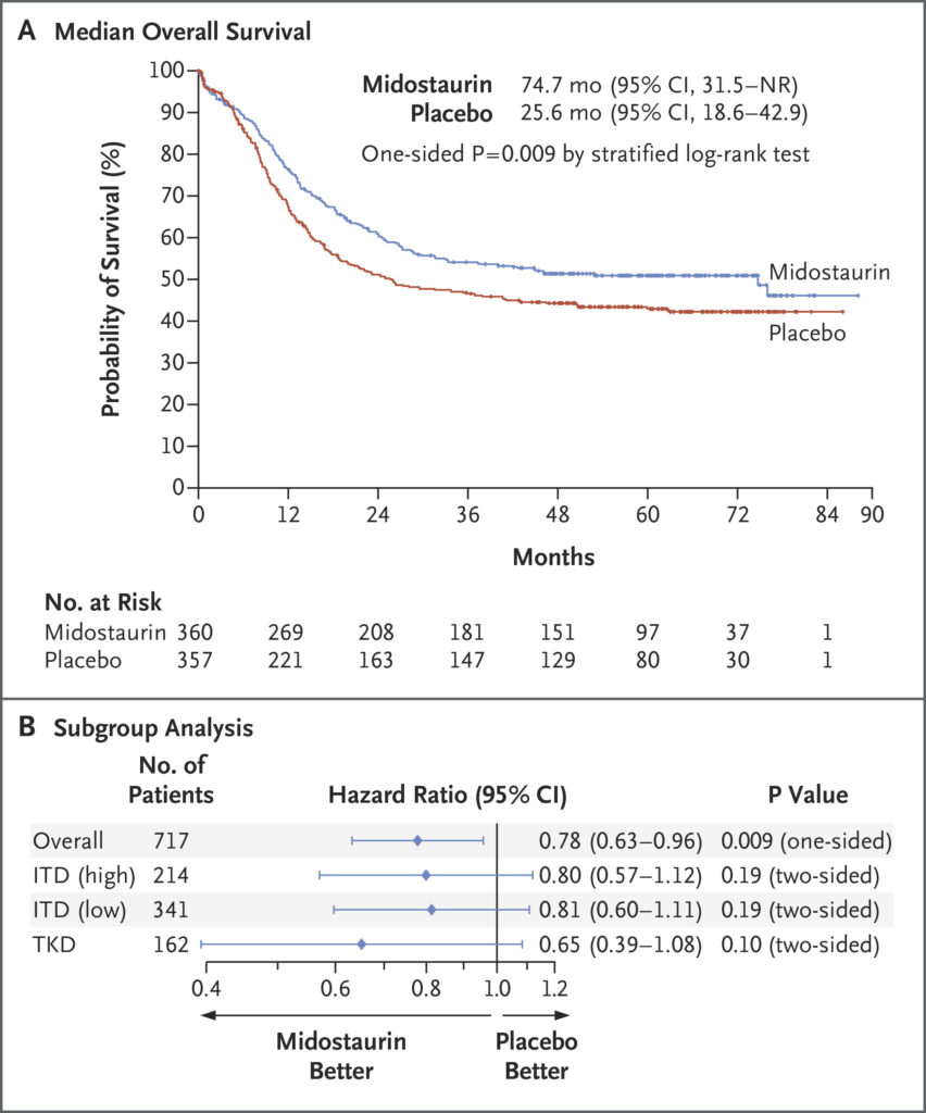
    • Overall Survival (OS): The addition of midostaurin significantly improved OS. The median OS was 74.7 monthsin the midostaurin group compared to 25.6 months in the placebo group. The hazard ratio (HR) for death was 0.78(one-sided p = 0.009), indicating a 22% reduction in the risk of death.
    • Event-Free Survival (EFS): Midostaurin also improved EFS, with a median of 8.2 months versus 3.0 months in the placebo group (HR = 0.79; p = 0.0067).
    Kaplan-Meier survival curves from the RATIFY trial of midostaurin vs placebo in combination with intensive chemotherapy.

    https://www.nejm.org/doi/full/10.1056/NEJMoa1614359

    Long-Term Follow-Up

    A 10-year follow-up analysis confirmed the durability of midostaurin’s benefit:

    • OS Benefit: The survival advantage persisted over a decade, with a sustained improvement in OS for patients receiving midostaurin.
    • EFS Benefit: The improvement in EFS was maintained, reinforcing the long-term efficacy of midostaurin in combination with chemotherapy.

    Subgroup Analyses

    Midostaurin’s benefit was observed across various FLT3 mutation subtypes:

    • FLT3-ITD: Patients with both high and low allelic ratios of FLT3-ITD mutations experienced improved outcomes.
    • FLT3-TKD: Patients with TKD mutations also benefited from midostaurin addition.

    Limitations

    • Age Restriction: The trial included only patients aged 18–59, limiting the generalizability of results to older populations.
    • Maintenance Therapy: The study included a maintenance phase with midostaurin, but the specific contribution of maintenance therapy to overall outcomes remains unclear.
    • Toxicity: While midostaurin was generally well-tolerated, grade 3 infections occurred in 52% of patients in the midostaurin group, comparable to the placebo group.  

    In summary, the RATIFY trial established the addition of midostaurin to standard chemotherapy as a new standard of care for younger adults with FLT3-mutated AML, demonstrating significant improvements in survival outcomes. However, considerations regarding its applicability to older patients and the role of maintenance therapy warrant further investigation.

  • Deep Vein Thrombosis (DVT)

    Deep Vein Thrombosis (DVT)

    Deep Vein Thrombosis (DVT) is a common and potentially life-threatening condition caused by the formation of a thrombus in a deep vein, usually in the lower limbs. Prompt recognition and management are essential to prevent complications such as pulmonary embolism (PE) and post-thrombotic syndrome.

    Risk Factors (Virchow’s Triad)

    DVT risk is explained by Virchow’s Triad, which includes:

    • Venous stasis: Immobility, prolonged travel, recent surgery, heart failure.
    • Endothelial injury: Trauma, surgery, indwelling catheters, infection.
    • Hypercoagulability: Malignancy, pregnancy, oral contraceptives, thrombophilias (e.g., Factor V Leiden, antiphospholipid syndrome).

    Clinical Features

    • Unilateral limb swelling (most commonly calf or thigh).
    • Pain and tenderness along the deep venous system.
    • Erythema and warmth of the affected limb.
    • Homan’s sign (pain on dorsiflexion of the foot) is unreliable.

    Diagnosis

    Clinical Scoring System: The Wells Score

    • Used to stratify patients into low, moderate, or high risk for DVT.
    • score of ≥2 suggests DVT is likely.

    Investigations

    • D-dimer: High negative predictive value; useful to rule out DVT in low-risk patients.
    • Compression ultrasound (CUS): First-line imaging modality to confirm DVT.
    • Venography: Rarely used but considered the gold standard.

    Management

    Anticoagulation

    • First-line: Direct oral anticoagulants (DOACs) such as rivaroxaban or apixaban.
    • Alternative: Low molecular weight heparin (LMWH) with warfarin (target INR 2-3).
    • Duration:
      • Provoked DVT (e.g., surgery, transient risk factor): 3-6 months.
      • Unprovoked DVT: At least 6 months, consider indefinite treatment if high recurrence risk.
      • Cancer-associated DVT: LMWH or DOACs for at least 6 months. Generally indefinite if ongoing active malignancy.

    Supportive Measures

    • Compression stockings: Reduce post-thrombotic syndrome risk.
    • Early ambulation: Encouraged over prolonged bed rest.
    • Inferior vena cava (IVC) filter: Considered in select patients with temporary contraindications to anticoagulation.

    Thrombophilia Testing

    Thrombophilia testing is not routinely indicated in most patients with DVT. It is generally not useful for guiding treatment decisions and should be avoided in patients with provoked DVT (e.g., recent surgery, immobilization, malignancy) as it does not change management.

    When to Consider Thrombophilia Testing?

    • Unprovoked DVT in a young patient (<50 years old).
    • Recurrent unprovoked venous thromboembolism (VTE).
    • Strong family history of VTE.
    • Unusual site thrombosis (e.g., cerebral, mesenteric, portal vein thrombosis).
    • Suspected antiphospholipid syndrome (e.g., recurrent pregnancy loss, arterial thrombosis).

    Testing should always be performed after completion of anticoagulation therapy, as acute thrombosis and anticoagulation can alter results.

    Complications

    • Pulmonary embolism (PE): Sudden dyspnoea, pleuritic chest pain, haemoptysis.
    • Post-thrombotic syndrome: Chronic pain, swelling, and skin changes.
    • Recurrent DVT: Requires prolonged anticoagulation in high-risk individuals.

    Conclusion

    DVT is a critical diagnosis that requires prompt assessment and anticoagulation to prevent complications. Understanding risk factors, clinical presentation, and management principles is essential for junior doctors. Thrombophilia testing is not necessary in most cases and should only be considered in select patients where it may impact clinical management.

    Want to learn more? Visit HaematologyForDoctors.com!

  • A brief introduction to Hodgkin Lymphoma

    A brief introduction to Hodgkin Lymphoma

    Hodgkin Lymphoma (HL) is a malignant lymphoma characterised by the presence of Reed-Sternberg cells within an inflammatory background. It is an important and potentially curable haematological malignancy, especially in younger patients.

    Epidemiology

    • Bimodal age distribution: Peaks in young adults (15-35 years) and older adults (>55 years).
    • Slight male predominance.
    • Risk factors: Epstein-Barr Virus (EBV) infection, family history, immunosuppression (e.g., HIV, post-transplant patients).

    Clinical Presentation

    • Painless lymphadenopathy (most commonly cervical, supraclavicular, or mediastinal).
    • B symptoms (fever >38°C, drenching night sweats, weight loss >10% in 6 months) indicate advanced disease.
    • Pruritus and alcohol-induced lymph node pain are less common but characteristic.
    • Splenomegaly, hepatomegaly, or organ involvement in advanced disease.

    Diagnosis

    • Lymph node biopsy (excisional preferred) showing Reed-Sternberg cells in a mixed inflammatory background.
    • Immunohistochemistry: CD30+ and CD15+.
    • Staging investigations:
      • CT/PET scan – Determines extent of disease.
      • Bone marrow biopsy – Not routinely required
      • Blood tests: ESR, LDH, FBC (may show anaemia, leukocytosis, eosinophilia).

    Staging (Ann Arbor Classification)

    • Stage I: Single lymph node region.
    • Stage II: Multiple lymph node regions on the same side of the diaphragm.
    • Stage III: Lymph nodes on both sides of the diaphragm.
    • Stage IV: Disseminated involvement (e.g., liver, bone marrow).

    Management

    • Early-stage disease (I-II): Further classified into favourable and unfavourable based on factors such as bulky disease, high ESR, extranodal involvement, and multiple nodal sites.
    • Advanced-Stage disease (IIB-IV)
    • Treatment options include chemotherapy and radiotherapy.
    • ABVD or escalated BEACOPP: depending on stage and fitness of patient.
    • Relapsed/refractory disease: Salvage chemotherapy, autologous stem cell transplant, and checkpoint inhibitors (e.g., pembrolizumab) for resistant cases.

    Prognosis

    • Highly curable, with >90% survival in early-stage disease.
    • Poorer prognosis with B symptoms, bulky disease, and extranodal involvement.

    Hodgkin Lymphoma is a curable malignancy with well-defined treatment strategies. Prompt diagnosis and staging are key to achieving excellent outcomes.

    Want to learn more? Visit HaematologyForDoctors.com!

  • Acute vs Chronic Leukaemia – A Brief Overview

    Acute vs Chronic Leukaemia – A Brief Overview

    Leukaemia is a group of malignancies affecting the bone marrow and blood, characterised by the uncontrolled proliferation of abnormal white blood cells. It is broadly classified into acute and chronic types, which differ significantly in presentation, pathophysiology, and management. This guide provides a structured overview to help junior doctors navigate the diagnosis and treatment of leukaemia.

    Classification of Leukaemia

    Leukaemias are divided based on cell lineage (myeloid vs lymphoid) and disease course (acute vs chronic):

    • Acute Myeloid Leukaemia (AML): Rapidly progressive cancer of myeloid progenitor cells.
    • Acute Lymphoblastic Leukaemia (ALL): Rapidly progressive cancer of lymphoid progenitor cells.
    • Chronic Myeloid Leukaemia (CML): Slow-growing cancer of myeloid cells.
    • Chronic Lymphocytic Leukaemia (CLL): Slow-growing cancer of lymphoid cells.

    Pathophysiology

    • Acute leukaemias arise from immature precursor cells (blasts), leading to uncontrolled proliferation and bone marrow failure.
    • Chronic leukaemias involve the accumulation of more mature but dysfunctional cells, leading to a slower disease progression.

    Clinical Presentation

    Acute Leukaemias (AML & ALL)

    • Rapid onset over weeks to months.
    • Symptoms of bone marrow failure, including anaemia (fatigue, pallor), neutropenia (infections), and thrombocytopenia (bleeding, bruising).
    • Systemic symptoms such as fever, weight loss, and night sweats.
    • ALL can involve the central nervous system (CNS), causing headaches, seizures, and neurological deficits.

    Chronic Leukaemias (CML & CLL)

    • Insidious onset over years, often detected incidentally on routine blood tests.
    • Fatigue, weight loss, and night sweats.
    • Lymphadenopathy and splenomegaly, especially in CLL.
    • CML may present with abdominal discomfort due to splenomegaly and a high white cell count.

    Investigations

    Acute Leukaemia

    • Full blood count (FBC): High or low white cell count with circulating blasts; anaemia and thrombocytopenia are common.
    • Blood film: Presence of blasts.
    • Bone marrow biopsy: Confirms diagnosis with >20% blasts.
    • Cytogenetics: Identifies translocations such as PML-RARA in acute promyelocytic leukaemia (APL).

    Chronic Leukaemia

    • FBC: Leukocytosis in CML; lymphocytosis in CLL.
    • Blood film: Smudge cells in CLL; left shift (immature myeloid cells) in CML.
    • Bone marrow biopsy: Hypercellular marrow with mature cells.
    • Cytogenetics: BCR-ABL fusion gene in CML.

    Management

    Acute Myeloid Leukaemia (AML)

    • Intensive chemotherapy with daunorubicin and cytarabine.
    • Bone marrow transplantation for high-risk or relapsed cases.

    Acute Lymphoblastic Leukaemia (ALL)

    • Multi-agent chemotherapy.
    • CNS prophylaxis with intrathecal methotrexate.
    • Bone marrow transplant for high-risk patients.

    Chronic Myeloid Leukaemia (CML)

    • First-line treatment with tyrosine kinase inhibitors (TKIs) such as imatinib or dasatinib.
    • Long-term suppression of disease with excellent survival rates.

    Chronic Lymphocytic Leukaemia (CLL)

    • Early-stage disease is often observed without treatment.
    • Symptomatic or high-risk disease is treated with targeted therapies like BTK inhibitors (ibrutinib) or BCL-2 inhibitors (venetoclax), often combined with monoclonal antibodies like rituximab or obinutuzumab.

    Prognosis

    • Acute leukaemias progress rapidly and require urgent treatment. Survival rates vary: AML has a 40-50% survival rate in younger patients but much lower in the elderly. ALL has a 70-90% survival rate in children but lower in adults.
    • Chronic leukaemias progress slowly and are often manageable for years. CML has an excellent prognosis with TKIs, with >90% five-year survival. CLL is highly variable, with some patients living decades without treatment.

    Key Takeaways for Junior Doctors

    • Acute leukaemias present with bone marrow failure and require urgent treatment.
    • Chronic leukaemias are often incidental findings and may not need immediate therapy.
    • Blasts in blood and bone marrow (>20%) suggest acute leukaemia.
    • CML is driven by BCR-ABL and treated with TKIs.
    • CLL is often indolent but requires treatment in high-risk cases.

    Leukaemia is a complex but fascinating field in haematology. Junior doctors play a vital role in recognising its presentation, initiating investigations, and understanding treatment pathways.

    Want to learn more? Join our in-depth haematology course at HaematologyForDoctors.com!

  • Chronic Lymphocytic Leukaemia (CLL) – A Junior Doctor’s Guide

    Chronic Lymphocytic Leukaemia (CLL) – A Junior Doctor’s Guide

    Chronic Lymphocytic Leukaemia (CLL) is the most common adult leukaemia in Western countries. It is a clonal disorder of mature B lymphocytes and has a highly variable clinical course, ranging from an indolent disease requiring no treatment to aggressive forms necessitating early intervention. This guide provides an overview of CLL for junior doctors rotating through haematology.

    Understanding CLL

    CLL is characterised by the accumulation of small, mature-looking but dysfunctional B lymphocytes in the blood, bone marrow, and lymphoid tissues. It is closely related to small lymphocytic lymphoma (SLL), with the distinction being that CLL primarily affects peripheral blood, whereas SLL involves lymphoid organs.

    Clinical Presentation

    CLL can be asymptomatic and detected incidentally on a routine full blood count. Symptomatic patients may present with:

    • Lymphadenopathy – Most common initial presentation.
    • Hepatosplenomegaly – Occurs in advanced disease.
    • Recurrent infections – Due to immune dysfunction and hypogammaglobulinaemia.
    • Fatigue & Weight Loss – B symptoms in more aggressive disease.
    • Autoimmune Complications – Autoimmune haemolytic anaemia (AIHA) and immune thrombocytopenia (ITP).

    Investigations

    • Full Blood Count (FBC) & Blood Film – Lymphocytosis (>5 × 10⁹/L) with small mature lymphocytes and smear (smudge) cells.
    • Flow Cytometry – Confirms clonal B-cell population with CD5, CD19, CD20 (dim), and CD23 positivity.
    • Bone Marrow Aspirate & Biopsy – Not always required but shows increased lymphocytes.
    • Cytogenetics & Molecular Testing – Prognostic markers:
      • FISH for del(13q), del(11q), del(17p) (p53 mutation – poor prognosis).
      • IGHV mutation status (mutated = better prognosis, unmutated = worse prognosis).
    • Immunoglobulin Levels – Assesses hypogammaglobulinaemia.

    Staging

    Two staging systems guide prognosis and treatment decisions:

    • Rai Staging (USA):
      • 0: Lymphocytosis only (low risk).
      • I-II: Lymphadenopathy and/or organomegaly (intermediate risk).
      • III-IV: Anaemia and/or thrombocytopenia (high risk).
    • Binet Staging (Europe):
      • A: <3 lymphoid areas involved.
      • B: ≥3 lymphoid areas involved.
      • C: Anaemia and/or thrombocytopenia.

    Management

    Management depends on disease stage, symptoms, and genetic markers. Many patients do not require immediate treatment and are monitored under a watch and wait approach.

    Supportive Care

    • Infection Prevention – Vaccination (e.g., pneumococcal, influenza), IV immunoglobulin for recurrent infections.
    • Autoimmune Cytopenias – Managed with steroids, rituximab, or immunosuppressants.
    • Blood Transfusions – Symptomatic anaemia or thrombocytopenia.

    Definitive Treatment

    1. Indications for Treatment
      • Symptomatic disease (B symptoms, bulky lymphadenopathy, organomegaly).
      • Progressive marrow failure (anaemia or thrombocytopenia).
      • Rapid lymphocyte doubling time (<6 months).
      • Severe autoimmune complications unresponsive to steroids.
    2. First-Line Therapy
      • Targeted Therapy (preferred):
        • Bruton’s tyrosine kinase (BTK) inhibitors: Ibrutinib, Acalabrutinib.
        • BCL-2 inhibitors: Venetoclax (often combined with anti-CD20 monoclonal antibodies like rituximab or obinutuzumab).
      • Chemoimmunotherapy (for selected fit patients with IGHV-mutated disease and no TP53 mutation):
        • FCR regimen (Fludarabine, Cyclophosphamide, Rituximab) – Younger patients.
        • Bendamustine + Rituximab – Older or frail patients.
        • Note this is (extremely) rarely used in the modern era
    3. Relapsed/Refractory CLL
      • BTK inhibitors (Ibrutinib, Acalabrutinib) or BCL-2 inhibitors (Venetoclax) if not used first-line.
      • PI3K inhibitors (e.g., Idelalisib) in selected cases.
      • Allogeneic stem cell transplantation for high-risk, refractory disease.

    Palliative Care

    • For elderly or frail patients with slow disease progression, best supportive care is often appropriate.
    • Symptom management with transfusions, steroids for autoimmune cytopenias, and infection prophylaxis.
    • Clear goals-of-care discussions, particularly in advanced disease.

    Complications to Watch For

    • Infections – Bacterial, viral (zoster), and fungal due to immunosuppression.
    • Tumour Lysis Syndrome (TLS) – Need to assess TLS risk and institute management as required when starting venetoclax.
    • Richter’s Transformation – Transformation to an aggressive lymphoma (diffuse large B-cell lymphoma); suspect in rapidly enlarging lymph nodes or new B symptoms.
    • Autoimmune Cytopenias – Haemolytic anaemia or thrombocytopenia.
    • Secondary Malignancies – Increased risk due to immune dysfunction and prior treatments.

    Prognosis and Follow-Up

    • Indolent cases may never require treatment and have a normal life expectancy.
    • Patients with TP53 mutations, del(17p), or unmutated IGHV have a poorer prognosis and require novel targeted therapies.
    • Regular follow-up includes monitoring blood counts, lymphadenopathy, and signs of transformation or progression.

    Key Takeaways for Junior Doctors

    1. CLL is often asymptomatic and detected incidentally.
    2. Flow cytometry confirms the diagnosis by demonstrating a clonal B-cell population.
    3. Many patients follow a watch and wait approach, with treatment initiated only when clinically necessary.
    4. Targeted therapies (BTK and BCL-2 inhibitors) have replaced chemotherapy for most patients.
    5. Recognising Richter’s transformation and autoimmune complications is crucial for prompt intervention.
    6. Palliative care should be considered for frail patients with advanced disease.

    CLL is a fascinating and heterogenous disease with a rapidly evolving treatment landscape. Junior doctors play an essential role in recognising its presentation, monitoring progression, and initiating appropriate management.


    Want to expand your haematology knowledge? Our course at HaematologyForDoctors.com covers real-world case discussions, treatment updates, and practical management strategies. Sign up today!

  • Chronic Myeloid Leukaemia (CML) – A Junior Doctor’s Guide

    Chronic Myeloid Leukaemia (CML) is a myeloproliferative disorder characterised by the uncontrolled proliferation of myeloid cells in the bone marrow. It is unique among leukaemias due to its strong association with a specific genetic abnormality, the Philadelphia chromosome (t(9;22)), which leads to the formation of the BCR-ABL fusion gene. This guide provides an overview of CML for junior doctors rotating through haematology.

    Understanding CML

    CML is a triphasic disease with distinct clinical phases:

    • Chronic Phase – Most patients are diagnosed in this phase, which is typically indolent and well-managed with targeted therapy.
    • Accelerated Phase – Increased disease activity with worsening cytopenias, rising white cell counts, and worsening symptoms.
    • Blast Crisis – Resembles acute leukaemia, with a poor prognosis unless treated aggressively.

    Clinical Presentation

    CML is often detected incidentally on routine blood tests. However, symptomatic patients may present with:

    • Fatigue and weight loss – Due to increased metabolic activity.
    • Splenomegaly – Leading to early satiety, left upper quadrant pain, or abdominal discomfort.
    • Night sweats and fever – Indicative of disease progression.
    • Bone pain – Due to increased myeloid proliferation.
    • Bleeding/bruising – If thrombocytopenia develops in later stages.

    Investigations

    A structured diagnostic approach is key when CML is suspected:

    • Full Blood Count (FBC) & Blood Film – Shows leucocytosis with a left shift (neutrophils, myelocytes, basophilia, and eosinophilia).
    • Bone Marrow Aspirate & Biopsy – Hypercellular marrow with increased granulopoiesis.
    • Cytogenetics & Molecular Testing –
      • Philadelphia chromosome detection via karyotyping.
      • BCR-ABL1 fusion gene confirmed via PCR or fluorescence in situ hybridisation (FISH).
    • Lactate Dehydrogenase (LDH) & Uric Acid – Markers of high cell turnover.

    Management

    Treatment of CML has been revolutionised by the introduction of tyrosine kinase inhibitors (TKIs), which specifically target the BCR-ABL fusion protein.

    Supportive Care

    • Hydroxyurea – Used for cytoreduction in patients with very high white cell counts at diagnosis.
    • Allopurinol & Hydration – To prevent tumour lysis syndrome (TLS).

    Definitive Treatment

    1. Tyrosine Kinase Inhibitors (TKIs) – The cornerstone of CML treatment, including:
      • First-line: Imatinib (first-generation), Dasatinib, Nilotinib (second-generation).
      • For resistant disease: Bosutinib, Ponatinib (third-generation).
      • TKI response monitoring: Measured via BCR-ABL1 transcript levels using quantitative PCR.
    2. Stem Cell Transplantation – Considered for TKI-resistant disease or blast crisis.
    3. Chemotherapy – Used in blast crisis to induce remission before transplant.

    Palliative Care

    • For patients who fail multiple lines of therapy or are unfit for aggressive treatment, supportive care focuses on symptom management and maintaining quality of life.
    • Palliative options include hydroxyurea for cytoreduction and symptom control.
    • End-of-life discussions and advanced care planning should be initiated when disease progresses despite therapy.

    Complications to Watch For

    • Disease Progression – Transition from chronic to accelerated phase or blast crisis.
    • TKI Resistance or Intolerance – Requires switching therapy or alternative treatment strategies.
    • Cardiotoxicity & Vascular Events – Seen with second- and third-generation TKIs.
    • Myelosuppression – TKI-induced cytopenias may require dose modifications.
    • Tumour Lysis Syndrome (TLS) – Can occur at treatment initiation, requiring prophylactic measures.

    Prognosis and Follow-Up

    • With TKIs, CML has transformed into a manageable chronic condition with near-normal life expectancy if well-controlled.
    • Regular monitoring with BCR-ABL1 PCR ensures response to therapy and detects early resistance.
    • Some patients in deep remission may attempt treatment-free remission (TFR) under strict monitoring.

    Key Takeaways for Junior Doctors

    1. CML is strongly linked to the Philadelphia chromosome (t(9;22)) and BCR-ABL fusion gene.
    2. Tyrosine kinase inhibitors (TKIs) have revolutionised treatment and significantly improved survival.
    3. Monitoring BCR-ABL1 transcript levels via PCR is crucial for assessing treatment response.
    4. Recognising disease progression from chronic phase to blast crisis is critical.
    5. Palliative care is an important option for patients who are refractory to treatment or unfit for aggressive therapy.

    CML is a fascinating disease that exemplifies the power of targeted therapy in modern haematology. Understanding its phases, management, and complications will help junior doctors provide optimal patient care.


    Want to learn more about haematological malignancies? Our haematology course provides in-depth case discussions, management strategies, and exam-focused content. Sign up now at HaematologyForDoctors.com!

  • Acute Lymphoblastic Leukaemia (ALL) – A Junior Doctor’s Guide

    Acute Lymphoblastic Leukaemia (ALL) is an aggressive haematological malignancy resulting from the clonal proliferation of lymphoid precursor cells (blasts). It is more common in children but also occurs in adults, with prognosis and management differing by age group. Early recognition and timely intervention are key to improving patient outcomes. This guide provides an overview of ALL for junior doctors rotating through haematology.

    Understanding ALL

    ALL arises from malignant transformation of lymphoid progenitor cells, leading to uncontrolled proliferation and bone marrow failure. It is broadly classified into B-cell ALL (B-ALL) and T-cell ALL (T-ALL) based on the cell lineage of origin. Genetic and molecular features further stratify risk and guide treatment decisions.

    Clinical Presentation

    Patients with ALL often present with symptoms related to bone marrow failure and organ infiltration:

    • Anaemia – Fatigue, pallor, dyspnoea
    • Neutropenia – Recurrent infections, fever
    • Thrombocytopenia – Easy bruising, petechiae, mucosal bleeding
    • Lymphadenopathy & Hepatosplenomegaly – More common in T-ALL
    • Mediastinal Mass – T-ALL often presents with a mediastinal mass causing respiratory distress or superior vena cava syndrome (SVCS)
    • Neurological Symptoms – Headache, cranial nerve palsies, or seizures due to central nervous system (CNS) involvement

    Investigations

    A structured approach is crucial when suspecting ALL. Essential investigations include:

    • Full Blood Count (FBC) & Blood Film – Shows anaemia, thrombocytopenia, and lymphoblasts.
    • Bone Marrow Aspirate & Biopsy – Confirms the diagnosis with >20% lymphoblasts in the marrow.
    • Immunophenotyping (Flow Cytometry) – Differentiates B-ALL from T-ALL.
    • Cytogenetics & Molecular Testing – Identifies key mutations (e.g., Philadelphia chromosome t(9;22), TEL-AML1, MLL rearrangements) for prognosis and targeted therapy.
    • Lumbar Puncture – Evaluates for CNS involvement.
    • Coagulation Studies – Important in cases with disseminated intravascular coagulation (DIC).
    • Chest X-ray/CT – Checks for a mediastinal mass in T-ALL.

    Management

    Management of ALL is intensive and consists of several phases:

    Supportive Care

    • Blood Product Support – Red cell and platelet transfusions as needed.
    • Infection Control – Empirical broad-spectrum antibiotics for febrile neutropenia, antifungal and antiviral prophylaxis in high-risk cases.
    • Tumour Lysis Syndrome (TLS) Prophylaxis – Hydration, allopurinol, or rasburicase to prevent TLS.

    Definitive Treatment

    1. Induction Therapy (Aim: Achieve remission)
      • Multi-agent chemotherapy including vincristine, steroids (dexamethasone or prednisolone), anthracyclines (e.g., doxorubicin), and asparaginase.
      • CNS-directed therapy with intrathecal methotrexate.
    2. Consolidation (Intensification) Therapy (Aim: Eradicate residual disease)
      • High-dose methotrexate, cytarabine, and additional systemic chemotherapy.
      • Targeted therapies for specific subtypes (e.g., tyrosine kinase inhibitors [TKIs] for Philadelphia chromosome-positive ALL).
    3. Maintenance Therapy (Aim: Prevent relapse)
      • Long-term low-intensity chemotherapy (e.g., mercaptopurine, methotrexate) for 2-3 years, especially in paediatric protocols.
    4. Stem Cell Transplantation
      • Considered in high-risk or relapsed cases, particularly in adult ALL.
    5. Targeted Therapy
      • TKIs (e.g., imatinib, dasatinib) for Philadelphia chromosome-positive ALL.
      • Monoclonal antibodies (e.g., blinatumomab, inotuzumab) for relapsed or refractory disease.

    Palliative Care

    • In patients who are unfit for intensive therapy due to age, comorbidities, or frailty, supportive care focuses on symptom management and quality of life.
    • Transfusions for anaemia-related symptoms, infection control, and pain management are key.
    • Clear discussions about goals of care and advanced care planning should be undertaken.

    Complications to Watch For

    • Febrile Neutropenia – Requires urgent broad-spectrum antibiotics.
    • Tumour Lysis Syndrome (TLS) – Can lead to acute kidney injury and metabolic derangements.
    • Disseminated Intravascular Coagulation (DIC) – Common in hyperleukocytosis.
    • CNS Involvement – Requires intrathecal chemotherapy.
    • Relapsed/Refractory Disease – May require novel therapies or stem cell transplantation.

    Prognosis and Follow-Up

    • Prognosis is better in children, with cure rates exceeding 80%.
    • Adults have a lower survival rate, especially in high-risk subtypes.
    • Long-term follow-up includes:
      • Monitoring for late effects of chemotherapy (e.g., secondary malignancies, cardiotoxicity).
      • Surveillance for relapse with periodic blood tests and bone marrow biopsies.

    Key Takeaways for Junior Doctors

    1. Consider ALL in patients with pancytopenia and circulating lymphoblasts.
    2. Early initiation of supportive care, including infection control and TLS prophylaxis, is crucial.
    3. ALL management involves multiple chemotherapy phases and is highly protocol-driven.
    4. Recognising complications such as febrile neutropenia and mediastinal masses can be life-saving.
    5. Palliative care is an important consideration for patients unsuitable for intensive therapy.

    ALL is a complex but treatable disease, particularly with advances in targeted therapies and stem cell transplantation. Junior doctors play a key role in initial diagnosis, supportive care, and recognising complications.


    Want to gain a deeper understanding of haematological malignancies? Our comprehensive haematology course provides case-based learning, practical management strategies, and exam-focused content to help you excel. Sign up now at HaematologyForDoctors.com!

  • Acute Myeloid Leukaemia (AML) – A Junior Doctor’s Guide

    Acute Myeloid Leukaemia (AML) – A Junior Doctor’s Guide

    Acute Myeloid Leukaemia (AML) is a rapidly progressive haematological malignancy that originates from the myeloid lineage of blood cells. It is a crucial diagnosis to be aware of when working in haematology or oncology, as early recognition and management significantly impact patient outcomes. This guide provides a structured overview of AML for junior doctors rotating through a haematology service.

    Understanding AML

    AML is characterised by clonal proliferation of immature myeloid blasts in the bone marrow, leading to bone marrow failure and subsequent cytopenias. The condition is highly heterogeneous, with multiple subtypes classified based on genetic and molecular features. It is most commonly seen in older adults but can also affect younger patients.

    Clinical Presentation

    Patients with AML often present with symptoms related to bone marrow failure, including:

    • Anaemia – Fatigue, pallor, shortness of breath
    • Neutropenia – Increased susceptibility to infections, fever
    • Thrombocytopenia – Easy bruising, petechiae, mucosal bleeding
    • Systemic Symptoms – Weight loss, night sweats, malaise

    Some patients may present with complications such as leucostasis, which occurs due to a very high white cell count leading to symptoms of hyperviscosity, including headaches, confusion, and respiratory distress.

    Investigations

    A systematic approach is required when suspecting AML. Key investigations include:

    • Full Blood Count (FBC) & Blood Film – Typically shows anaemia, thrombocytopenia, and circulating blasts.
    • Bone Marrow Aspirate & Biopsy – Confirms the diagnosis with >20% blasts in the marrow.
    • Immunophenotyping (Flow Cytometry) – Differentiates AML from other leukaemias.
    • Cytogenetics & Molecular Studies – Essential for risk stratification and prognosis (e.g., FLT3, NPM1, CEBPA mutations).
    • Coagulation Profile – Especially important if DIC is suspected.
    • Lactate Dehydrogenase (LDH) & Uric Acid – Markers of high cell turnover.

    Management

    Management depends on the patient’s age, fitness, and genetic risk stratification.

    Supportive Care

    • Blood Product Support – Red cell transfusions for symptomatic anaemia, platelet transfusions to prevent bleeding.
    • Infection Control – Empirical antibiotics for febrile neutropenia, prophylactic antifungals and antivirals in high-risk patients.
    • Tumour Lysis Syndrome (TLS) Prophylaxis – Hydration, allopurinol, or rasburicase to prevent TLS.

    Definitive Treatment

    1. Intensive Chemotherapy
      • Induction Therapy – Standard 7+3 regimen (7 days of cytarabine + 3 days of an anthracycline such as daunorubicin)
      • Consolidation Therapy – High-dose cytarabine or allogeneic stem cell transplant in high-risk patients
    2. Low-Intensity Therapy
      • Hypomethylating agents (e.g., azacitidine, decitabine) for frail or elderly patients
      • Venetoclax-based regimens for patients who are unsuitable for intensive chemotherapy
    3. Targeted Therapy (based on molecular mutations)
      • FLT3 inhibitors (midostaurin, gilteritinib)
      • IDH1/IDH2 inhibitors (ivosidenib, enasidenib)
    4. Palliative Care
      • In patients who are unfit for treatment due to age, frailty, or comorbidities, the focus may shift to symptom management and quality of life.
      • Supportive measures include blood transfusions, pain management, and symptom control.
      • Discussions around goals of care and advanced care planning are essential in these cases.

    Complications to Watch For

    • Febrile Neutropenia – A medical emergency requiring broad-spectrum antibiotics.
    • Disseminated Intravascular Coagulation (DIC) – Seen in acute promyelocytic leukaemia (APL), requiring urgent all-trans retinoic acid (ATRA).
    • Leucostasis – High WBC counts causing microvascular obstruction, often requiring leukapheresis or hydroxyurea.
    • Relapsed/Refractory Disease – May require salvage chemotherapy or experimental therapies.

    Prognosis and Follow-Up

    AML prognosis depends on cytogenetic and molecular risk factors, patient age, and treatment response. Younger patients with favourable-risk AML can achieve long-term remission with intensive chemotherapy and stem cell transplantation. However, older patients or those with high-risk mutations often have poorer outcomes.

    After treatment, patients require regular follow-up with:

    • FBC monitoring for relapse detection
    • Bone marrow biopsies post-treatment
    • Long-term surveillance for complications such as secondary malignancies and graft-versus-host disease in transplant recipients

    Key Takeaways for Junior Doctors

    1. Always suspect AML in a patient with pancytopenia and circulating blasts.
    2. Early bone marrow biopsy and cytogenetic testing are crucial for diagnosis and treatment planning.
    3. Supportive care, including infection control and transfusions, is vital in management.
    4. Recognising complications such as febrile neutropenia and DIC can be life-saving.
    5. AML treatment is increasingly personalised, with targeted therapies improving outcomes.
    6. Palliation is an important option for patients who are unfit for treatment, with a focus on quality of life.

    AML is a challenging but fascinating disease that junior doctors will frequently encounter in haematology. Understanding its presentation, workup, and management will enable you to provide better care for your patients and work effectively within the haematology team.


    Want to learn more? Our comprehensive haematology course provides in-depth case-based learning, practical management tips, and exam-focused content to help you excel during your haematology rotation. Sign up now at HaematologyForDoctors.com!